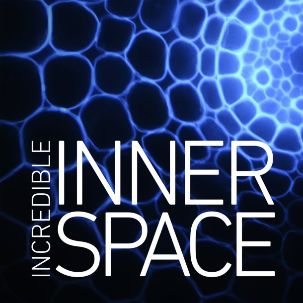
The Incredible Inner Space exhibition currently on display at the Embassy of Australia in Washington, D.C. presents the micro patterns in nature and innovative material with an unexpected result, art.
Developed by the Australian Microscopy and Microanalysis Research Facility (AMMRF), the traveling photography exhibition reveals the human quest for knowledge and curiosity of structures, space, and perspective.
Each image engages the viewer to inquire “What is this?” As a result, it becomes an opportunity to discover nature’s complex designs and patterns.
Incredible Inner Space is an innovative initiative to bring new technology in health and food sciences to the public. The collection of photographs is stunning with bold colors, delicate textures, familiar architectures, and classic organic relationships. Yet, the Incredible Inner Space exhibition is in reality a journey through cells, tissue, bone, and nano technology within material. Combining beautiful and intriguing visuals with knowledge is powerful.
Incredible Inner Space is engaging and appealing; and, it brilliantly demonstrates art in science.
By Keri Douglas, founder/editor 9MusesNews.com. © 2017. All Rights Reserved.
The Incredible Inner Space is on exhibit at the Embassy of Australia until July 2017. (Free and open to the public at Gallery at the Embassy of Australia, 1601 Massachusetts Avenue, NW, Washington, DC 20036, 10-2 pm, M-F)
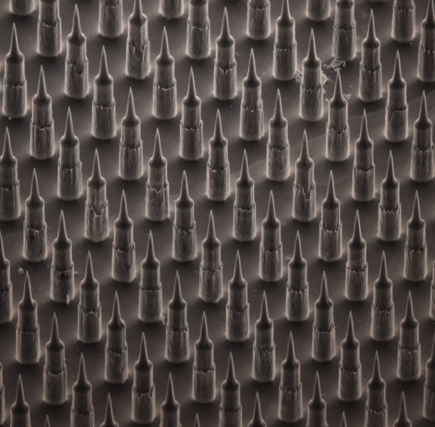
Can you guess what this is? This photo is of the fabric of a patch designed to deliver medication evenly through a multitude of tiny pin pricks. Imagine the potential.
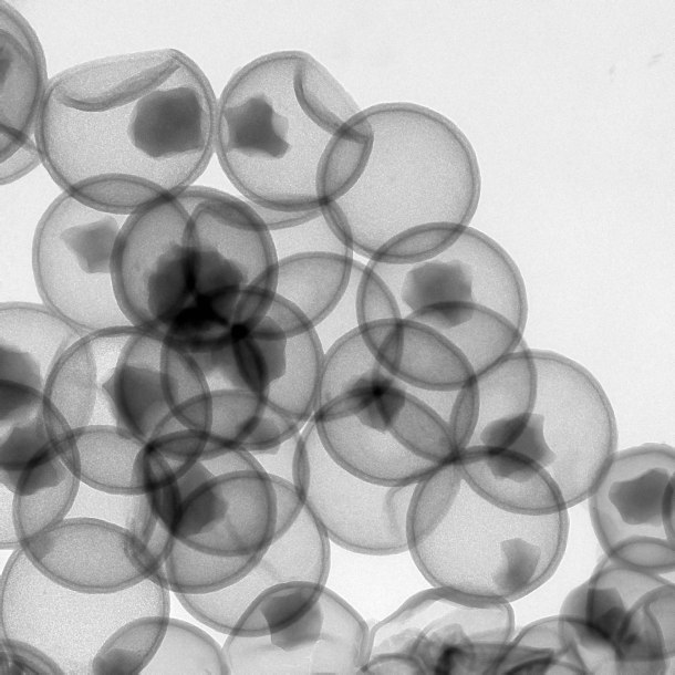
Date: 8 July 2011 Technique: FEI Tecnai G2 20 TEM Sample: Silica hollow sphere for drug delivery Scale: eg: horizontal field width is 500 nm What does the image show? Hollow silica nanoparticles were prepared from separate but concurrent core-dissolution and shell-growth processes, which occur when solid silica spheres were mixed with sodium borohydride. Transmission electron microscopy allows the process for forming the hollow spheres to be monitored. Here we can see that most of the hollow sphere still contain a smaller core particles, which indicate that the core-dissolution has not proceed to completion.

HFW= 216microns

Some thing new and informative!
https://creativegustorebel.wordpress.com follow up mine
Thanks for posting this! I definitely want to go to this exhibit!